
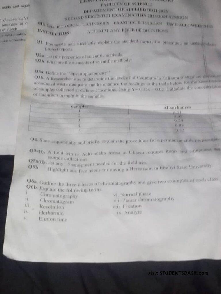
BIO 206 Past Questions and Answers

Join @Studentsdash on TG Assignment Solved by Studentsdash 1a. In your own way define management. 1b….
Join @Studentsdash on TG
Join @Studentsdash on TG ENG-207 Past questions and answers
Join @Studentsdash on TG
Join @Studentsdash on TG BUA 106: Introduction to Financial Accounting Assignment Questions and Answers Question Impact…
Join @Studentsdash on TG